

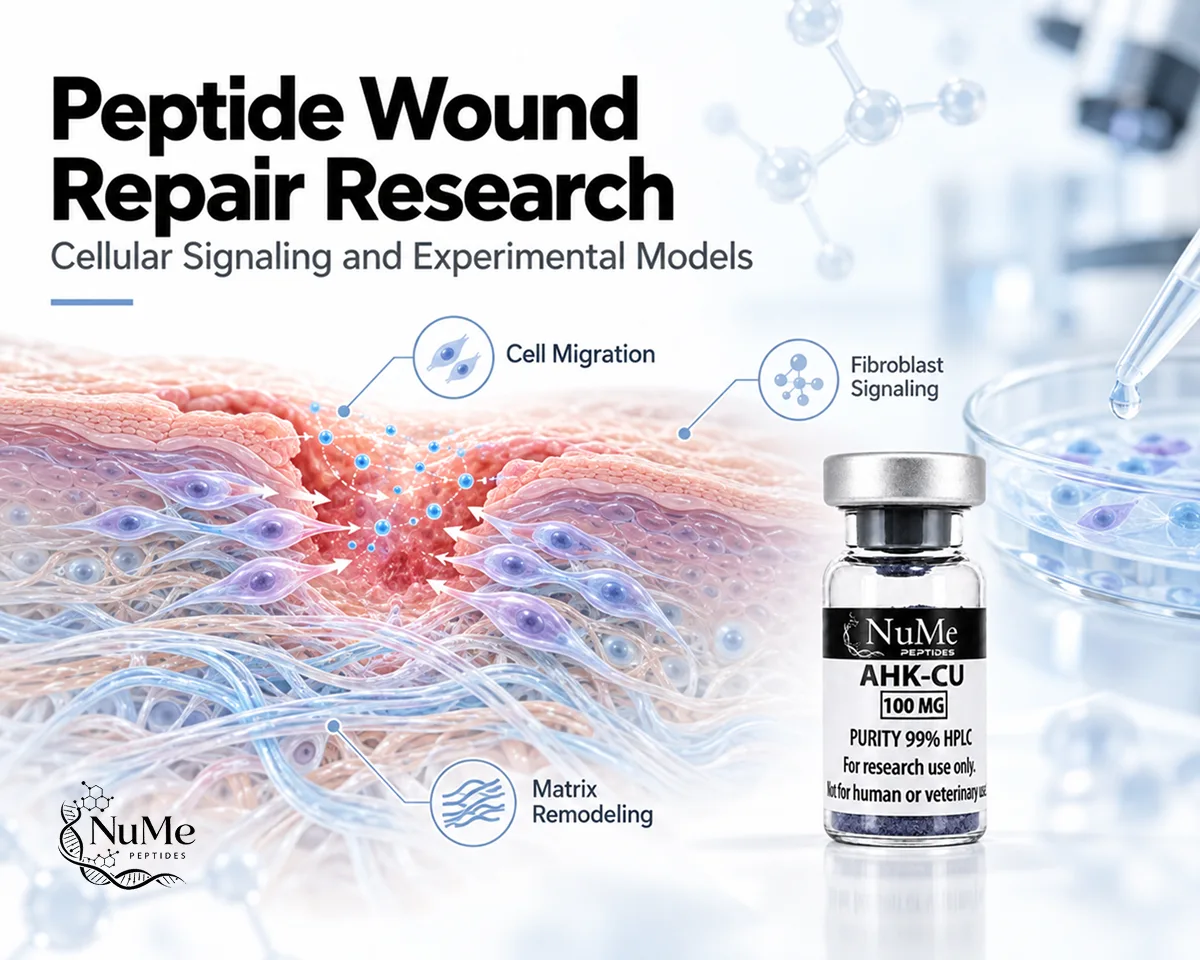
Peptide Wound Repair Research: Cellular Signaling and Experimental Models


Wound repair is a coordinated biological response involving platelets, immune cells, keratinocytes, fibroblasts, endothelial cells, extracellular matrix proteins, and multiple signaling pathways. Following tissue injury, these systems must work in sequence while also overlapping in time to limit damage, rebuild the wound bed, restore surface integrity, and reorganize newly formed tissue.
Interest in peptide wound repair research focuses on whether specific peptide molecules can influence particular stages of this process, including cell migration, angiogenesis, granulation-tissue formation, extracellular matrix turnover, and collagen organization. BPC-157 and thymosin beta-4-related compounds such as TB-500 are frequently discussed in this field, although their evidence bases are not equivalent and remain largely preclinical.
Understanding these compounds requires examining the exact experimental model and biological endpoint rather than treating “wound healing” as one measurable event.
Explore the NuMe Peptides US Shop to review research-focused peptide materials for laboratory and scientific investigation.
How Is Wound Repair Studied in Scientific Research?
Researchers study wound repair by following biological changes from the moment tissue is damaged through later structural remodeling.
The process is generally divided into overlapping phases: hemostasis, inflammation, proliferation, and remodeling. Hemostasis limits blood loss and creates an initial fibrin-based matrix. Inflammatory cells then enter the injured region, remove damaged material, and release signals that influence later repair. During proliferation, keratinocytes, fibroblasts, and endothelial cells become highly active, while remodeling gradually reorganizes extracellular matrix and collagen architecture. (Guo & DiPietro, 2010)
Experimental models are designed to isolate different parts of this sequence.
For example, cell-culture systems can measure keratinocyte or fibroblast migration. Endothelial assays can evaluate vascular behavior. Animal wound models provide a more complete environment in which inflammation, angiogenesis, matrix formation, and re-epithelialization occur together.
Common wound models include:
- full-thickness skin excisions;
- incisional wounds;
- burns;
- chemical injuries;
- ischemic wounds;
- diabetic wound models;
- implanted scaffold or sponge models.
Researchers may then measure wound area, re-epithelialization, granulation tissue, vascular density, collagen deposition, inflammatory markers, matrix-remodeling enzymes, or mechanical strength.
Because each measurement represents only one aspect of repair, stronger experimental studies typically combine several endpoints.
Explore research compounds studied in tissue repair and regeneration through our Healing & Recovery collection.
Which Cellular Stages Are Involved in Wound Repair?
Wound repair progresses through overlapping biological stages rather than discrete steps that completely stop before the next one begins.
Hemostasis
Immediately after tissue disruption, platelets aggregate and fibrin forms an initial clot.
This clot limits bleeding but also functions as a provisional matrix through which cells later migrate. Platelets release signaling molecules that help recruit inflammatory and reparative cell populations.
Inflammation
Neutrophils and macrophages enter the wound and participate in debris removal, antimicrobial defense, and signaling.
Appropriate inflammatory activity is necessary, but persistent inflammation can delay the transition toward proliferation and tissue reconstruction. Chronic wounds are often characterized by prolonged inflammatory signaling and impaired responses from epidermal and dermal cells. (Broughton et al., 2012)
Proliferation
The proliferative phase involves several major cellular events.
Keratinocytes migrate across the wound surface to re-establish the epithelial barrier. Fibroblasts proliferate and deposit extracellular matrix. Endothelial cells contribute to new vessel formation, while granulation tissue develops within the wound bed.
A detailed review of acute wound biology describes this phase as a combination of fibroplasia, re-epithelialization, angiogenesis, and tissue restoration involving multiple interacting cell types. (Gonzalez et al., 2016)
Remodeling
Later, the initially formed matrix undergoes structural modification.
Collagen fibers become reorganized, matrix components are degraded and replaced, and cellularity often decreases as the wound matures.
These overlapping phases explain why researchers evaluating experimental peptides need to identify which stage and cell population the compound actually affects.
What Role Do Cell Migration and Matrix Remodeling Play in Research?
Cell migration is essential because wound closure requires several cell populations to move into or across damaged tissue.
Keratinocytes at the wound edge adopt a more migratory phenotype and move laterally across the wound surface during re-epithelialization. Fibroblasts migrate into the wound bed, where they contribute new extracellular matrix and may later differentiate into myofibroblasts. Endothelial cells migrate during angiogenesis to establish vascular support. (Wilkinson & Hardman, 2020)
Thymosin beta-4 provides an experimental example of how peptide research can focus directly on migration.
In human endothelial-cell models, thymosin beta-4 increased directional migration and accelerated movement across scratch-wounded endothelial monolayers. The study also reported increased matrix metalloproteinase production, linking cell movement with modification of the surrounding matrix.
Matrix remodeling is equally important.
The extracellular matrix must provide temporary structural support while remaining dynamic enough for cells to migrate and tissue to mature. Matrix metalloproteinases, or MMPs, break down specific matrix proteins, allowing older or provisional components to be removed.
Thymosin beta-4 has been shown to increase several MMPs during wound repair. Mouse wound experiments demonstrated increased MMP-2 and MMP-9 expression, while cultured keratinocytes, endothelial cells, fibroblasts, and monocytes showed cell-specific changes in MMP-1, MMP-2, and MMP-9. (Philp et al., 2006)
This illustrates why wound-repair studies often measure migration and matrix turnover together.
Explore research compounds studied in tissue repair and regeneration through our Healing & Recovery collection.
How Are Peptides Investigated in Wound Repair Models?
Researchers typically investigate peptides by matching an experimental compound with a defined wound model and a set of measurable outcomes.
One study may focus on endothelial migration. Another may examine epithelial closure, granulation-tissue formation, collagen deposition, or matrix remodeling.
Cell Migration Assays
Scratch assays and Boyden chamber systems allow researchers to measure whether a peptide changes the movement of keratinocytes, fibroblasts, or endothelial cells.
Thymosin beta-4 has been particularly well studied using this type of model. A rat full-thickness wound study found increased keratinocyte migration in vitro along with enhanced re-epithelialization, angiogenesis, collagen deposition, and wound contraction in vivo. (Malinda et al., 1999)
Vascular Models
Researchers may measure endothelial proliferation, VEGF expression, or vascular tube formation because angiogenesis helps supply the wound bed with oxygen and nutrients.
BPC-157 has been investigated through this approach. In a rat alkali-burn wound model, BPC-157 was associated with increased wound closure, granulation tissue, re-epithelialization, dermal remodeling, and collagen deposition. Complementary endothelial experiments showed increased proliferation, migration, VEGF expression, and vascular tube formation involving ERK1/2-related signaling. (Huang et al., 2015)
Histology and Structural Assessment
Animal tissue sections can be evaluated for collagen arrangement, vascularity, inflammatory cells, epithelial coverage, and granulation tissue.
These measurements provide greater biological context than cell assays alone.
Mechanical Testing
For incisional or mechanically active wounds, tensile strength may also be assessed.
Early BPC-157 experiments used rat skin incisions, colon anastomoses, and implanted sponges to study collagen, blood-vessel formation, granulation tissue, and tensile strength. (Sikiric et al., 1997)
Together, these approaches help distinguish a molecular response from a broader wound-repair phenotype.
Which Research Peptides Are Studied in Wound Repair Research?
BPC-157
BPC-157 is a synthetic 15-amino-acid peptide investigated predominantly in preclinical wound and tissue-injury models.
One of the more directly relevant studies used an alkali-burn model of skin injury. BPC-157-treated animals showed greater wound closure, improved granulation-tissue formation, increased re-epithelialization, dermal remodeling, and collagen deposition. In complementary endothelial experiments, the peptide increased VEGF-related signaling, migration, proliferation, and vascular tube formation.
Earlier animal experiments also examined multiple repair endpoints rather than wound closure alone.
Skin incisions, colon anastomoses, and implanted sponge models were used to assess angiogenesis, collagen formation, granulation tissue, and tensile strength.
Despite this preclinical research base, evidence in humans remains extremely limited.
A systematic review published in October 2026 identified only three eligible human BPC-157 studies involving 30 total participants. The authors rated overall certainty of evidence as very low because of methodological weaknesses, indirectness, and imprecision. (Poolton et al., 2026)
Therefore, BPC-157 is best described as an experimental peptide with preclinical wound-repair evidence rather than an established human wound treatment.
TB-500 and Thymosin Beta-4
The evidence base for thymosin beta-4 is more extensive than direct evidence specifically generated with TB-500.
Thymosin beta-4 is a naturally occurring 43-amino-acid peptide associated with actin regulation. Research has identified active regions involved in cell migration, angiogenesis, wound repair, survival, and inflammatory regulation.
In a classic full-thickness rat wound model, thymosin beta-4 increased re-epithelialization, wound contraction, collagen deposition, and angiogenesis. It also produced a strong increase in keratinocyte migration in vitro. (Malinda et al., 1999)
Additional research has linked thymosin beta-4 with extracellular matrix remodeling through increased MMP expression during the early stages of wound repair. (Philp et al., 2006)
TB-500 is commonly discussed as a thymosin beta-4-related peptide, but researchers should confirm the exact molecule used in each study before transferring conclusions from native thymosin beta-4 to TB-500.
A recent orthopaedic and regenerative review similarly concluded that TB-4 and TB-500 have promising preclinical evidence related to angiogenesis and tissue repair, while robust human orthopaedic evidence remains lacking.
FAQs About Peptide Wound Repair Research
What is wound repair research?
It examines the cellular and molecular processes that occur after tissue injury, including inflammation, keratinocyte migration, fibroblast activity, angiogenesis, extracellular matrix deposition, and remodeling.
Why is cell migration important during wound repair?
Keratinocytes must migrate to restore the epithelial barrier, fibroblasts move into the wound bed to produce matrix, and endothelial cells migrate during vascular formation.
How is BPC-157 studied in wound models?
Preclinical studies have examined burn healing, granulation tissue, re-epithelialization, collagen deposition, angiogenesis, endothelial migration, and vascular signaling.
How is thymosin beta-4 connected with wound repair?
Experimental studies have reported effects involving keratinocyte and endothelial migration, angiogenesis, collagen deposition, re-epithelialization, and matrix metalloproteinases.
Can thymosin beta-4 evidence automatically be applied to TB-500?
No. They are closely related in current peptide discussions, but researchers should identify which molecular form was actually investigated before attributing findings to TB-500.
Do animal wound studies establish human effectiveness?
No. Cell and animal studies help define mechanisms and generate hypotheses, but human safety and efficacy require appropriately controlled clinical research.
Understanding peptide wound repair research requires examining the wound as a changing biological environment rather than reducing repair to how quickly an injury closes.
Hemostasis establishes the initial provisional matrix. Inflammation clears damaged material and coordinates signaling. Keratinocytes, fibroblasts, and endothelial cells then migrate and proliferate, while later matrix remodeling reorganizes collagen and other structural components.
BPC-157 has been investigated in preclinical wound models involving angiogenesis, granulation tissue, re-epithelialization, and collagen deposition. Thymosin beta-4 research provides particularly direct evidence involving cellular migration, vascular development, extracellular matrix enzymes, and wound closure, while TB-500-specific conclusions require greater care because much of the foundational evidence comes from native thymosin beta-4.
The most informative studies therefore combine molecular signaling with cell behavior, tissue histology, matrix organization, and functional outcomes.
Explore the NuMe Peptides US Shop to review research-focused peptide materials for laboratory and scientific investigation.
Disclaimer: This content is provided for scientific and educational purposes only. Research peptides discussed here are presented in a laboratory research context and are not described as products for human consumption, diagnosis, treatment, cure, or prevention of disease.